Recently, the joint research of Artificial Intelligence Assisted System for Cancer Bone Scintigraphy were accomplished by the researchers from the Department of Nuclear Medicine of West China Hospital and the College of Computer Science of Sichuan University, and the outcomes were published in Medical Image Analysis (IF: 11.5), a top journal in the interdisciplinary field of nuclear medicine and AI. This is also the largest single-center research about the application of AI in nuclear medical bone imaging analysis.
Single photon emission computed tomography (SPECT) full body bone imaging (hereinafter referred to as “bone scanning” or BS) refers important value in the diagnosis of clinical tumor bone metastasis, and is also the most frequently utilized diagnostic technology in clinical nuclear medicine. A recent Chinese national nuclear medicine annual census showed that more than 2.1 million SPECT examinations were carried out every year in this county, of which the bone imaging accounts for 61.3%, i.e. the number of annual examinations exceeds 1.25 million. By now, WCH serves the largest number of bone scanning patients in China. In 2019, 43000 patients were examined, with an average of over 130 cases per day, which occupies an enormous workload for SPECT physicians. However, the limited resolution of BS images makes the interpretation is time-consuming and experience-dependent work and has the disadvantages of subjectivity, error distinctive, and unsatisfied efficiency.
Recently, the development of artificial intelligence (AI) is creeping into every facet in modern life by its advances in big-data retrieval and explicit feature evaluation, which is ideal for medical image analysis. Compared with PET/CT or PET/MR images, SPECT BS planar images contain relative less characteristic image features, or, from another perspective, the complexity of imaging features were simpler, which is beneficial for image feature extraction and AI model construction.
Thus, the researchers from the Department of Nuclear Medicine of WCH and the Medical AI Center of the College of Computer Science, SCU, carried out the application research on the specific clinical topic of “AI assisted diagnosis of tumor bone metastasis.”
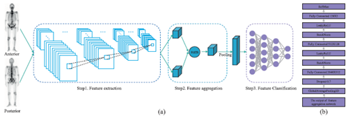
The study included 15,474 full-body bone images of tumor patients completed in the Department of Nuclear Medicine of WCH from 2018 to 2019. With the application of the deep convolutional neural networks and the spatial attention feature fusion operator technology, an AI model was successfully constructed for bone imaging-assisted diagnosis. The comprehensive diagnostic accuracy of bone metastasis was 93.17% for 22 types of cancers, of which the successful judgment for lung cancer, prostate cancer and breast cancer was above 95%. This is helpful to better incorporate the limited medical human resources into the diagnosis and treatment of difficult and critical diseases, and render good prospects for clinical application.
At present, the research team of Nuclear Medicine has developed a new type of AI assisted diagnostic system based on this technology. Its prototype system has been installed in the Department for closed beta tests. It is expected to be successfully applied and promoted into clinic in the future.